NANOSYSTEMS: PHYSICS, CHEMISTRY, MATHEMATICS, 2018, 9 (1), P. 120–122
New superparamagnetic fluorescent Fe@C-C5ON2H10-Alexa Fluor 647 nanoparticles for biological applications
A. S. Garanina – GREMAN, UMR CNRS 7347, Francois Rabelais University, Tours, France; National University of Science and Technology “MISiS”, Moscow, Russia
I. I. Kireev – Belozersky Institute of Physico-Chemical Biology, Moscow State University, Moscow, Russia
I. B. Alieva – Belozersky Institute of Physico-Chemical Biology, Moscow State University, Moscow, Russia
A. G. Majouga – National University of Science and Technology “MISiS”, Moscow; Faculty of Chemistry, Lomonosov Moscow State University, Moscow, Russia
V. A. Davydov – L. F. Vereshchagin Institute for High Pressure Physics of the RAS, Moscow, Russia
S. Murugesan – Center for Technology Innovation, Baker Hughes a GE Company, Houston, TX, USA
V. N. Khabashesku – Center for Technology Innovation, Baker Hughes a GE Company, Houston, TX, USA
V. N. Agafonov – GREMAN, UMR CNRS 7347, Francois Rabelais University, Tours, France
R. E. Uzbekov – Faculty of Medicine, Francois Rabelais University, Tours, France; Faculty of Bioengineering and Bioinformatics, Moscow State University, Moscow, Russia; rustem.uzbekov@univtours.fr
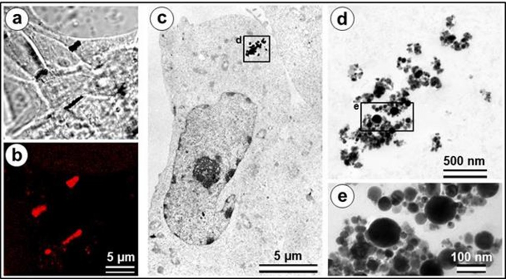
The structure and physical properties of superparamagnetic Fe@C nanoparticles (Fe@C NPs) as well as their uptake by living cells and behavior inside the cell were investigated. Magnetic capacity of Fe@C NPs was compared with Fe7C3@C NPs investigated in our previous work, and showed higher value of magnetic saturation, 75 emu.g-1 (75 Am2•kg-1), against 54 emu.g-1 (54 Am2•kg-1) for Fe7C3@C. The surface of Fe@C NPs was alkylcarboxylated and further aminated for covalent linking to the molecules of fluorochrome Alexa Fluor 647. Fluorescent Fe@C-C5ON2H10-Alexa Fluor 647 NPs (Fe@C-Alexa NPs) were incubated with HT1080 human fibrosarcoma cells and investigated using fluorescent, confocal laser scanning and transmission electron microscopy. No toxic effect on the cell physiology was observed. In a magnetic field, the NPs became aligned along the magnetic lines inside the cells.
Keywords: superparamagnetic fluorescent Fe@C nanoparticles, electron microscopy, magnetic field.
PACS 68.37.Lp, 75.75.Cd
DOI 10.17586/2220-8054-2018-9-1-120-122
