NANOSYSTEMS: PHYSICS, CHEMISTRY, MATHEMATICS, 2019, 10 (2), P. 215–226
Electron microscopy of biogenic minerals: structure and sizes of uranium dioxide nanoparticles with Mn2+ impurities
E. I. Suvorova – A.V. Shubnikov Institute of Crystallography, Federal Scientific Research Centre “Crystallography and Photonics” of Russian Academy of Sciences, Leninsky pr., 59, 19333 Moscow, Russia; suvorova@crys.ras.ru
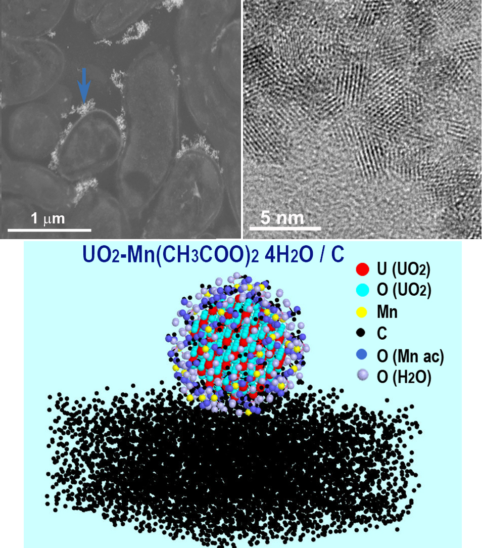
Methodological aspects of the extraction of structural and chemical information from transmission electron microscopy (TEM) of uranium dioxide (UO2) biogenic nanoparticles are presented. Nanoparticles were formed via the bacterial reduction of water-soluble uranyl acetate with U (VI) in the presence of Mn2+ ions and cultures Shewanella oneidensis MR-1 in the medium. The particles of 1.2 – 3.5 nm in diameter and particle agglomerations were visualized in conventional TEM, high resolution TEM, scanning TEM modes. Their phase and chemical composition were investigated with electron diffraction, X-ray energy dispersive spectrometry and electron energy loss spectroscopy with high spatial resolution. Maintenance of the element balance helped to find the composition of the mixture of UO2 and Mn acetates. The interpretation of TEM data and modeling allowed to propose the mechanism for the suppression of UO2 particle growth and higher resistance to dissolution of smaller UO2 particles with adsorbed Mn acetate compared to the larger pure particles.
Keywords: uranium dioxide nanoparticles, bacterial reduction, manganese impurity, transmission electron microscopy.
PACS 61.46.Df, 68.37.Lp, 87.17.-d
DOI 10.17586/2220-8054-2019-10-2-215-226
